 Understanding MSK
Medullary Sponge Kidney · Rein en éponge médullaire
A rare diagnosis, explained with care.
Found after years of stones and infections, MSK quickly hits a wall of jargon. Here, in plain language, is what we know — and everything that exists elsewhere in the world.
For the people who live with MSK — and the doctors they show this page to. Every fact, a link to its source.
What is MSK?
A condition present from birth, usually benign.
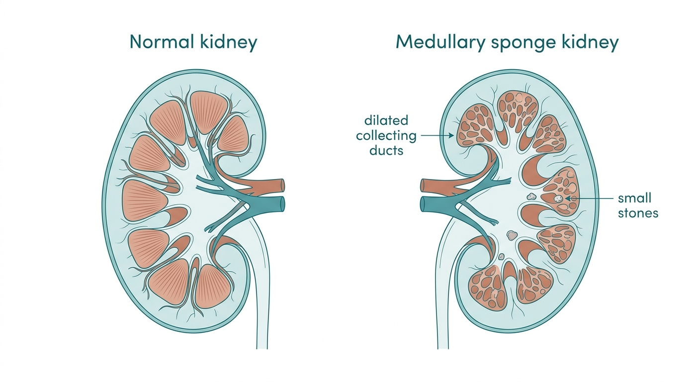
In medullary sponge kidney, the small collecting ducts in the core of the kidney (the medulla) are dilated, giving a "sponge-like" appearance. This is present from birth and is generally benign for kidney function.
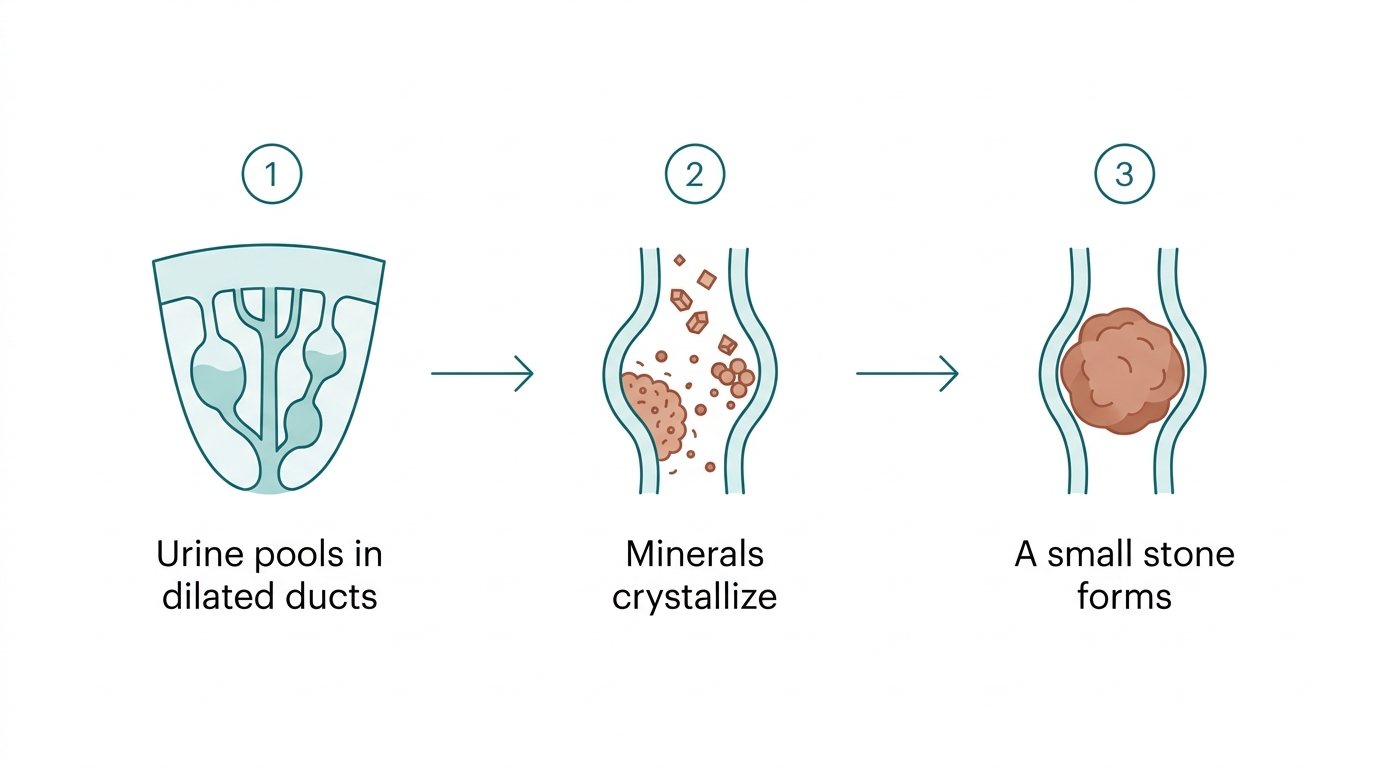
The trouble comes from stasis: urine pools in these widened ducts, which favours stone formation, urinary infections and sometimes blood in the urine. That’s often how it’s discovered, between ages 20 and 40. MSK accounts for 12–20% of people who form recurrent calcium stones.
Sources: NIDDK StatPearls
Recognizing the signs
How MSK shows up.
Recurrent stones
the most common sign
Blood in urine
visible or microscopic
Urinary infections
sometimes recurrent
Flank pain
acute or chronic
Often silent
found by chance
Epidemiology
Rare doesn’t mean imaginary.
MSK affects roughly 1 in 5,000 people — often silent, it is almost certainly under-counted. But in the stone clinic, it is far more common than people think.
the estimated prevalence — but up to 30% among young women who form stones. The first stone often appears between 15 and 36.
How a stone forms
From stasis to stone, in three steps.
Stone composition
MSK stones aren’t just oxalate stones.
This is the point your doctor will want to see: MSK stones lean strongly toward calcium phosphate, a sign of a kidney acidification defect. Prevention depends on it.
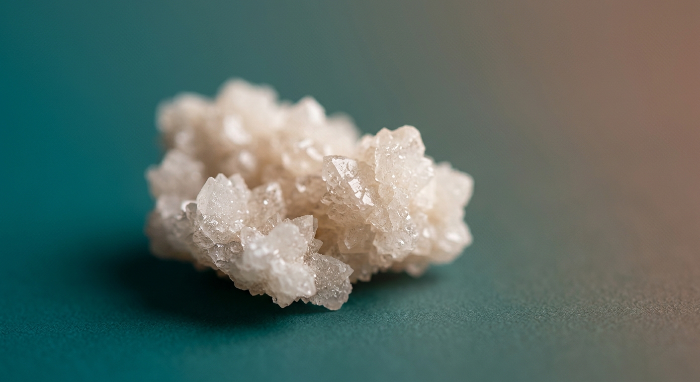
MSK vs non-MSK — Daudon 2022, 1,036 stones
The IVa2 morphology — a sign of distal renal tubular acidosis — is 30× more common in MSK. C. R. Chimie 2022
General population — German registry, 45,783 analyses
~80% of stones contain calcium. World J Urol 2022
What works
The interventions, from least to most invasive.
Each option with its level of evidence, real numbers and source. The most useful step isn’t always surgery.
The best-proven prevention in MSK
Long-term potassium citrate cut stone events from 0.58 to 0.10 stones per year in MSK patients — and also reduced ureteroscopies, lithotripsies and hospitalizations.
Potassium citrate
Prevention — best-proven in MSK
A daily alkali salt that raises urinary citrate (a natural stone inhibitor) and lowers urinary calcium.
Hydration + low-sodium diet
The foundation, no real risk
Drinking enough to make ~2 L of urine a day dilutes the urine and limits crystal formation. A lifelong baseline for MSK patients who form stones.
Thiazide diuretics
Questioned since 2023
Lower urinary calcium. Long prescribed, but the randomized NOSTONE trial (2023, 416 patients) showed no clear benefit vs placebo in the general population. May still help when hypercalciuria dominates — worth discussing.
Medical expulsive therapy (tamsulosin)
Helps pass a stone
An alpha-blocker that relaxes the ureter to help a distal stone ≤10 mm pass. Strong AUA recommendation.
Shockwave lithotripsy (ESWL)
Least invasive
Shockwaves break the stone from outside the body, no incision. Less effective for large (>2 cm) or very dense stones.
Laser ureteroscopy (URS)
Often first-line
A thin scope travels up the urinary tract and a laser fragments or "dusts" the stone. Higher stone-free rate than ESWL depending on size and location.
Thulium-fiber laser (TFL)
Beat holmium in recent RCTs
A newer laser than holmium:YAG. In a randomized trial, markedly higher stone-free rate for kidney stones.
Percutaneous nephrolithotomy (PCNL / mini-PCNL)
For large stones
Direct access to the kidney through a small incision in the back. Best stone-free rate for large volumes, but the most invasive. Mini-PCNL lowers complications.
Stone culture (not urine alone)
Safety — specific to MSK
Because MSK stones often harbour bacteria, culturing the removed stone predicts post-op sepsis better than a routine urine culture.
Renal denervation (refractory pain)
Under study — not standard care
Interrupting the kidney’s nerves for severe, refractory chronic flank pain. Published, but limited to case series — not an established treatment.
Urease / anti-biofilm microrobots
Preclinical — research
Magnetic microrobots that chemically dissolve stones or disperse the bacterial biofilm. Promising, but lab-only for now.
Supplement to discuss
Kidney C.O.P.: worth evaluating, not proof by itself.
Kidney C.O.P. means Calcium Oxalate Protector. It is a U.S. supplement aimed at calcium oxalate stones. It can be brought to an appointment, but it does not replace a urine work-up, stone analysis, or an individualized medical plan.
What the product contains
According to the NIH label: serving size is 2 delayed-release vegetarian capsules.
An honest read of the evidence
The formula is patented and the manufacturer highlights laboratory inhibition of calcium oxalate crystals. That is biologically interesting, but it is not the same as a clinical trial showing fewer stones in MSK patients.
Status
U.S. dietary supplement — not a prescription medication.
Claimed target
Calcium oxalate crystals and stones.
Product evidence
Mostly lab / patent / in-vitro support; no strong human clinical trial proof found that it prevents or dissolves stones.
U.S. usage
No reliable public user count found. Store reviews and testimonials are not usage data.
Patient reviews
Official testimonials are positive; retail reviews are mixed, with some reporting benefit and others no effect.
MSK / hospital review
No Memorial Sloan Kettering monograph specific to Kidney C.O.P. found in quick search.
Official Kidney C.O.P. site 2024 review — alkalinizing agents
Ask the doctor
The useful question is not only “is it good?”
The useful question is: which stone type and urine profile are we trying to correct? MSK can involve calcium oxalate, calcium phosphate, low citrate, higher urine pH or other drivers. The choice depends on the work-up.
- Are the stones confirmed calcium oxalate, calcium phosphate, mixed, or another type?
- Should a 24-hour urine profile be done before choosing a supplement?
- Is there low citrate, high calcium, high oxalate, high pH, or low urine volume?
- Would prescribed potassium citrate be more appropriate than this supplement?
- Is this product safe with current medications, history, and kidney function?
- Should vitamin C supplements be avoided while focusing on hydration, lower sodium, and normal dietary calcium?
National Kidney Foundation — calcium stones Johns Hopkins — kidney stones
Diet
Your fork and your water glass: your first medicine.
Because MSK stones are mostly calcium-based, food and hydration are the most powerful, lowest-risk levers. A starting point to bring to a dietitian.
Do
- Drink steadily all day — aim for ~2.5 L of urine (≈12–13 cups), sipped like a slow river.
- Get calcium from food (~1,000–1,200 mg/day, with meals) — it binds oxalate.
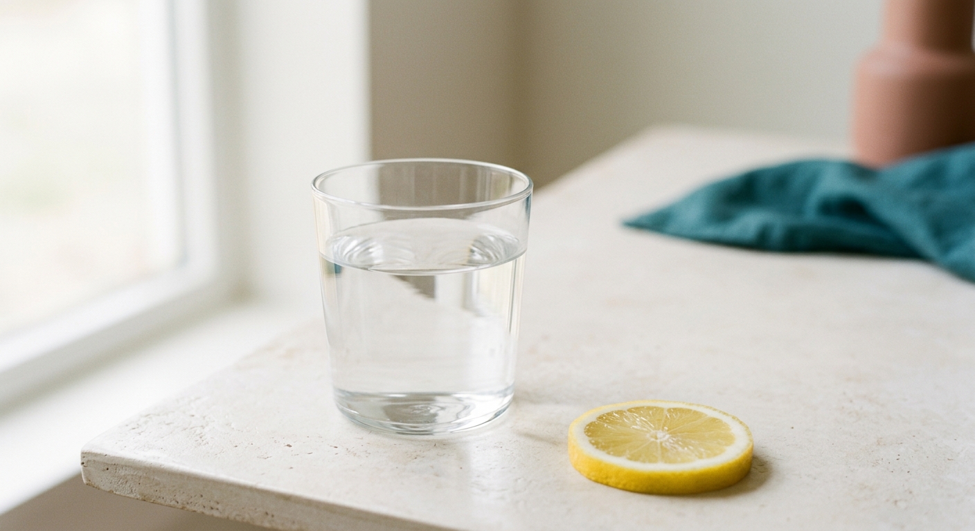
- Add a daily squeeze of lemon or lime to water (citrate + pH).
- Fill the plate with fruits and vegetables — at least 5 servings a day.
- Replace some meat with plant protein (beans, peas, lentils).
Limit
- Sodium — under 2,300 mg/day: high salt pushes calcium into the urine.
- Non-dairy animal protein (meat, organ meats, eggs, shellfish).
- Sugary drinks and excess juice.
- Vitamin C supplements (they convert to oxalate).
- Don’t cut dietary calcium — a low-calcium diet raises stone risk (~+50%).
A normal-calcium, low-sodium, low-animal-protein diet cut recurrence roughly in half (RR 0.49). Borghi et al., NEJM 2002
Looking ahead
Today’s research, tomorrow’s treatments.
Real leads, presented honestly: promising research, no promises. MSK has no cure — but the field is finally moving, and fast.
The latest · 2025–2026
Each entry verified to its primary source (PMID / DOI confirmed).
Exome sequencing in MSK
Genetic heterogeneity confirmed — no single gene (Lyon cohort).
Renal denervation & MSK pain
First dedicated review: a lead for refractory chronic pain.
Bacterial biofilms inside stones
Biofilms are intrinsic internal components of calcium stones.
Deep phenotyping: BOLD-MRI + measured GFR
Impaired medullary oxygenation; eGFR over-reads kidney function.
“Porous perspectives” — major MSK review
The most up-to-date comprehensive synthesis.
The leads that could change things
Chronic pain finally recognized
Since 2018, the literature recognizes MSK chronic pain as its own syndrome — sometimes without an active stone. Treatment research is beginning.
Targeting the biofilm inside the stone
Bacteria live in nearly 40% of stones. Disrupting this biofilm (chitosan, 2025) could one day reduce infections and recurrences.
Stone-dissolving microrobots
Magnetic urease microrobots chemically dissolve stones in the lab. Promising, still far from the clinic.
Genetics: toward personalized medicine
Exome sequencing reveals variants in some patients — a step toward personalized follow-up of severe or familial forms.
Outlook & associated conditions
A lifelong companion — not a sentence.
Most kidneys keep their function for life. Serious decline is the exception and comes from complications, not the "sponge" itself. But a few things are worth a watchful eye.
keep normal kidney function for life
incomplete distal RTA (worth screening)
osteopenia — often improved by citrate
progress to kidney failure (complicated cases, upper bound)
J Nephrology 2025 (measured GFR) CJASN — bone & citrate Cleveland Clinic
The patients · living with MSK
“What I needed most was to be believed.”
For a minority, MSK means years of unexplained pain and the exhausting work of being taken seriously. These lines, gathered from patient communities, capture what comes up most.
Your pain is real. You are not faking it — even on the days the scans look quiet.
Rare doesn’t mean imaginary. About one in five thousand people carries these kidneys — you are uncommon, not unbelievable.
The big picture is kind: most kidneys with this stay strong for a whole lifetime. The work is in the watching, not the worrying.
What I needed most wasn’t a cure that day. It was one doctor who took me seriously.
Non-identifying themes from a patient-community audit + J Nephrology 2018 — MSK chronic pain
Québec vs the world
Where Québec is strong, where it differs.
Honestly: Québec is not an MSK research hub (Italy is), but its public coverage and official French-language guideline are real strengths.
| Dimension | Québec | Elsewhere in the world |
|---|---|---|
| Is a cure possible? | No — lifelong care of complications | No, anywhere (NIDDK: "scientists have not found a cure") |
| Reference guideline | CUA 2022 — official French version downloadable | AUA (US), EAU 2024 (Europe), NICE NG118 2019 (UK) |
| MSK research hub | Not a recognized hub | Italy leads — disease described in Padua; Verona centres |
| Coverage / access | Strength: single-payer public system | US: private insurance; UK/Europe: public, variable |
| Stone composition analysis | INESSS: recognized and recommended | Recommended everywhere (AUA, EAU, NICE, CUA) |
| MSK-specific prevention (citrate) | A concrete option to discuss with your doctor | Citrate listed by all societies; MSK evidence came from Italy |
Year of the latest guideline
The AUA medical guideline (2014) is the oldest; the CUA (2022) exists in French.
The Italian legacy
MSK carries the name Lenarduzzi–Cacchi–Ricci, described in Padua in the 1930s. To this day, the largest cohorts and the prevention evidence (citrate) come from Italy — Verona above all.
Where the expertise is
From Montréal to Verona, the map of knowledge.
MSK is treated close to home — and the deepest research comes from Italy. Real centres, with their links, from Québec to the historic cradle of the disease.
Québec
- CHUM — Nephrology & UrologyMontréal — Stone metabolic work-up, endourology · 514-890-8000
- MUHC / McGill — Stone disease (Dr. Sero Andonian)Montréal — Recognized in minimally invasive stone surgery, ESWL
- CHU de Québec–Université Laval — UrologyQuébec City — Urinary stones, ureteroscopy, PCNL (referral required)
- RI-MUHC — Metabolic Disorders (MeDiC)Montréal — Kidney & metabolic disease research
Canada
- Stone Centre — Vancouver General HospitalVancouver — Canada’s first dedicated stone centre (Dr. Ben Chew)
- St. Michael’s — Kidney Stone Prevention ClinicToronto — Metabolic prevention: nephrologist + dietitian
- The Ottawa Hospital — Lithotripsy UnitOttawa — Extracorporeal shockwave lithotripsy (ESWL)
United States
- Rare Kidney Stone Consortium — Mayo ClinicRochester, MN — Rare stone diseases (Dr. Dawn Milliner)
- UCLA UrologyLos Angeles — 2026 discovery of bacterial biofilms inside stones
- Stanford UrologyStanford, CA — Chitosan / biofilm-dispersal research (2025)
Europe
- Hôpital Tenon (AP-HP) — CRISTALParis — Highly specialized urinary-stone centre
- Hôpital Édouard-Herriot (HCL)Lyon — MSK MRI phenotyping (Tournebize team)
- ERKNet — European rare kidney disease network24 countries — Network of reference centres
Italy — the cradle
- University of Verona — NephrologyVerona — World MSK hub: Prof. Giovanni Gambaro, Dr. Antonia Fabris
- University of Padua — NephrologyPadua — MSK genetics & cell biology (Prof. Franca Anglani)
- Policlinico Gemelli — Catholic UniversityRome — Stone research (Dr. Pietro Manuel Ferraro)
United Kingdom
- Guy’s & St Thomas’ — Kidney Stone UnitLondon — ESWL, surgery, cystinuria clinic · 020 7188 7638
- UCLH — Stones & EndourologyLondon — Specialist service + quantitative urine analysis
- Cambridge University Hospitals — Metabolic ClinicCambridge — Metabolic clinic (rare disease incl. stones)
Resources and support
Organizations that walk alongside you.
To learn, ask questions, and not feel alone.
To bring to your doctor
The questions that move the appointment forward.
A list ready to print or show — to cover the essentials without forgetting anything.
- Is the MSK diagnosis confirmed by imaging, and is it unilateral or bilateral?
- Is there nephrocalcinosis, discrete stones, or both?
- When was the last complete 24-hour urine profile?
- What were the values: volume, citrate, calcium, oxalate, sodium, urate, pH, creatinine?
- Has a stone ever been chemically analyzed? What type (oxalate, phosphate, struvite, mixed)?
- Is potassium citrate indicated or already optimized?
- Is hypercalciuria present, and would a thiazide be appropriate — despite NOSTONE?
- What daily urine-output target should I aim for?
- Is real stone-burden being tracked (not just "stable")?
- Would a volumetric CT comparison help?
- Is kidney function measured precisely enough for MSK (measured GFR, MRI)?
- If infections recur despite negative cultures, could stones be acting as a bacterial reservoir?
- If a stone is removed, can it be sent for chemical analysis AND culture?
- Before ESWL, ureteroscopy or PCNL, what is the antibiotic and culture plan?
- Would a stone-clinic (nephrology) opinion in addition to urology help?
- Is nephrology genetics relevant in my case?
- Can MSK cause chronic pain without visible obstruction, and what is the evaluation plan?
- What symptoms require emergency care?
When to seek emergency care
These signs can indicate a serious infection or an obstruction. They warrant a visit to the emergency room:
- Fever with flank pain
- Suspected urinary obstruction
- Signs of sepsis (chills, confusion, low blood pressure)
- Uncontrollable pain
- Persistent vomiting
- Drop in urine output
The science in depth — for nephrologists and urologists.
Ten precise facts, each linked to its primary source. The technical layer most MSK pages leave out — meant to be read, and checked, in consultation.
- 01
Plugs, not plaque. The only systematic intraoperative biopsy (n=12): MSK stones are mostly intratubular ("marbles" inside dilated collecting ducts); Randall’s plaque exceeded normal in only 6/12. MSK is a ductal-plugging disease, not a plaque disease.
- 02
The "tissue ossification" model falsified in vivo. Bone-gene / mineral co-localization in just 1 of 34 areas (P<0.001), and none in the ducts where stones actually form.
- 03
~14.5% of sporadic MSK patients (8/55, P=0.023) carry rare non-coding GDNF variants, absent in 170 control chromosomes, with 0 RET mutations — the ureteric-bud developmental axis.
- 04
"Sporadic" MSK is often familial. Screening 50 probands found 59 affected relatives across 27 families (autosomal dominant, reduced penetrance) — a reason to image first-degree relatives.
- 05
Only ~25% (4/15) of patients labeled "MSK" actually met imaging criteria — widespread over-diagnosis since non-contrast CT replaced urography.
- 06
eGFR over-reads kidney function in MSK. Iohexol-measured GFR 78 vs 90 mL/min/1.73m² (p=0.008) while eGFR showed no difference — a normal creatinine can falsely reassure.
- 07
The furosemide+fludrocortisone test over-diagnoses incomplete dRTA: specificity only 24% vs NH₄Cl (39% of "abnormals" were normal on the reference test).
- 08
A 0.35-unit rise in urine pH doubles the risk of a calcium-phosphate vs oxalate stone — over-alkalinizing to chase citrate can turn an oxalate former into a phosphate former.
- 09
Potassium citrate improved bone, not just stones: lumbar T-score −2.82 → −1.98 (P<0.001), alongside an ~83% drop in stone rate.
- 10
NOSTONE (NEJM 2023): hydrochlorothiazide did not prevent recurrence (placebo 59% vs 49% at 50 mg, NS) — but it never tested citrate/alkali, the mainstay of MSK. No verdict against potassium citrate.
The evidence
Everything verified, everything linked.
Every claim was cross-checked and its link tested. Research drew on NIDDK, INESSS, NICE, EAU, AUA, CUA and peer-reviewed studies, then verified link by link.